Discover what liver elastography is, how it works, and why it’s a valuable tool for assessing liver health without invasive procedures.
Many people experience unexplained fatigue, abdominal discomfort, or elevated liver enzymes during routine blood work. These symptoms can be concerning, often prompting doctors to investigate the health of the liver. Traditionally, diagnosing liver conditions and assessing their severity involved invasive procedures like liver biopsies. However, advancements in medical technology have introduced less intrusive methods that provide valuable insights. One such method is liver elastography, offering a clearer picture of liver health with greater comfort for patients.
What is Liver Elastography?
Liver elastography is a non-invasive diagnostic technique used to measure the stiffness of liver tissue. This stiffness, or elasticity, is directly related to the amount of scar tissue (fibrosis) or fat (steatosis) present in the liver. As liver diseases progress, the liver becomes more fibrotic and less elastic, essentially hardening.
Think of it like squeezing a sponge versus a brick. A healthy liver is more pliable, like a wet sponge, while a severely scarred liver is rigid and unyielding, like a brick. Elastography quantifies this difference, helping doctors understand the stage of liver disease and guide treatment decisions.
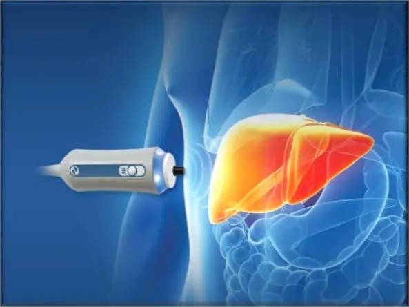
How Does Liver Elastography Work?
The core principle behind liver elastography is to send vibrations or ultrasound waves into the liver and measure how quickly they travel through the tissue. Denser, stiffer tissue transmits these waves faster than healthy, pliable tissue. The device then translates these measurements into a numerical value, often displayed as kilopascals (kPa) or meters per second (m/s).
Different types of elastography exist, but they all aim to achieve the same goal: assessing liver stiffness without the need for a biopsy. This makes it a significantly more comfortable and safer option for many patients.
Key Concepts Defined
- Fibrosis: The formation of excess fibrous connective tissue in the liver, a common response to chronic liver injury.
- Cirrhosis: Advanced fibrosis where the liver tissue is extensively scarred and damaged, impairing its function.
- Steatosis: The accumulation of fat in the liver, often associated with metabolic syndrome and obesity.
- Shear Wave Velocity: The speed at which shear waves propagate through the liver tissue, directly correlating with stiffness.
A Deeper Look at Liver Elastography Methods
While the goal is the same, there are a few primary methods used for liver elastography. Understanding these can help demystify the process further.
Transient Elastography (TE)
This is one of the most widely used methods. It employs a specific ultrasound probe that delivers a tiny, painless vibration to the skin over the liver. The ultrasound waves then measure the speed of the shear waves generated by this vibration as they pass through the liver. The results are typically displayed as a stiffness value in kPa.
- How it’s performed: The patient lies on their back, and the probe is placed on the skin between the ribs. The measurement takes only a few minutes.
- What it measures: Primarily fibrosis, but some devices can also assess fat content.
Magnetic Resonance Elastography (MRE)
MRE combines MRI technology with mechanical waves. A special pad is placed on the abdomen to generate low-frequency waves. The MRI scanner then creates detailed images of the liver, mapping the propagation of these waves. This method provides a 3D map of liver stiffness.
- How it’s performed: Requires an MRI scanner, so it’s typically done in a radiology suite. The patient lies inside the MRI machine.
- What it measures: Excellent for assessing both fibrosis and steatosis, and can cover a larger volume of the liver compared to some ultrasound-based methods.
Point Shear Wave Elastography (pSWE)
Similar to transient elastography, pSWE uses ultrasound but generates its own shear waves by focusing ultrasound pulses. It then measures the speed of these waves. This method can provide real-time stiffness measurements at specific points within the liver.
- How it’s performed: Performed using a standard ultrasound machine with elastography capabilities.
- What it measures: Provides quantitative stiffness measurements, often displayed in kPa.
Common Mistakes and Misconceptions
Even with advanced technology, understanding the nuances of liver elastography is crucial for accurate interpretation.
- Mistake: Assuming a single normal reading means no liver disease. While elastography is excellent, it’s a snapshot. Some conditions might not be fully apparent or might fluctuate.
- Misconception: Elastography replaces all liver biopsies. For certain complex cases or when definitive histological grading is absolutely necessary, a biopsy might still be considered.
- Mistake: Not considering other factors. Elastography results should always be interpreted alongside clinical history, blood tests, and imaging.
- Misconception: Any technician can perform it accurately. Proper technique and operator experience are vital for obtaining reliable results.
- Mistake: Ignoring the limitations of the specific device. Different elastography machines have varying sensitivities and specificities for different liver conditions and stages.
Practical Takeaways for Patients
If your doctor suggests a liver assessment, knowing about liver elastography test can empower you to ask the right questions. This non-invasive approach offers significant advantages:
- Comfort: It avoids the pain and risks associated with a liver biopsy.
- Speed: Measurements are usually quick, often taking less than 15 minutes.
- Accessibility: Increasingly available in many gastroenterology and radiology practices.
- Monitoring: Ideal for tracking the progression or improvement of liver disease over time.
A Decision-Making Perspective
From a patient’s viewpoint, the decision to undergo liver elastography is often straightforward. It represents a less daunting path to understanding liver health. For healthcare providers, it’s a valuable tool that helps differentiate between healthy liver tissue and fibrotic or fatty changes, guiding the need for further investigation or treatment initiation. It allows for more proactive management of chronic liver conditions, potentially preventing the development of serious complications like cirrhosis and liver failure.
Looking Ahead: The Evolving Role of Elastography
The field of liver elastography continues to evolve. Researchers are constantly working on improving the accuracy and expanding the capabilities of these technologies. Future developments may include even more precise assessments of different types of liver damage and better integration with artificial intelligence for more sophisticated analysis. As our understanding of liver diseases deepens, non-invasive tools like elastography will undoubtedly play an even more central role in patient care and management.
Also Read-The Science of Sole: How Footwear Affects Your Health